
The eye is one of the most complicated organs of the body, perhaps, the most complicated. Composed of many intricate parts, the eye bends light rays to stimulate nerve cells in the retina and by way of the optic nerve, send messages for interpretation by the brain. The brain uses the information from both optic nerves, and combines it to create one, clear image.
The connection between the eyes and brain is the reason for headaches when your vision changes and a loss of your peripheral vision due to stroke or tumor.
The eye can be divided into two segments: anterior and posterior segments.
The front of the eye: anterior segment

The anterior part of the eye includes the cornea, iris, ciliary body, and lens.
Cornea
The cornea is the clear dome on the surface of the eye. Its job is to focus light on the retina. It is the part of the eye that your contact lenses sit on. The cornea is also the most sensitive part of the eye. The cornea is dense with nerves and contains 70 to 80 large nerves and approximately 7,000 free epithelial nerve endings (nociceptors) per 1mm. The corneal nerves are responsible for the sensations of touch, pain, and temperature and play a role in blinking, healing, and tear production and secretion.
The cornea is covered by a tear film layer which recoats your eyes with each blink. The tear film layer of the eye performs many functions including forming tears and preventing evaporation of the tears.
The cornea can be affected by dry eye, red eyes and contact lens problems.
Iris
The iris is the colored part of the eye.
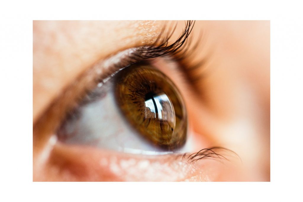
Did you know brown is the most common iris color? There are two muscles in the iris which allow the pupil to expand and contract. The iris allows light in and out of the eye through the pupil. Light sensitivity and difficulty with driving at night can be related to your pupil size; those with large pupils will have more halos and blur around car lights.
Ciliary body
The ciliary body is located towards the middle of the eye and performs two important functions. It produces a fluid called aqueous humor which maintains eye pressure and provides nutrients to the eye. Secondly, the ciliary body contains a ciliary muscle that controls the lens in the eye called accommodation.
Contraction and relaxation of this muscle and the lens is called accommodation. Accommodation is another word for focusing. This muscle allows you to see up close until the age of 40 until the muscle and lens structurally change.
Change in the ciliary muscle causes everyone to need some type of near correction for reading at some point in their lives.
Lens
The lens is located in the middle of the eye and is a biconcave, clear structure that helps the cornea focus light on the retina. The lens in the eye may become cloudy over time and result in blurry vision; a cloudy lens is called a cataract.
Cataracts are due to UV exposure from the sun or from long-term use of prescription medications like steroids. There are several types of cataracts that grow at different rates. Cataracts can cause blurry vision, more halo at night, and a yellowing tint to colors.
The back of the eye: posterior segment
The posterior part of the eye includes the retina, vitreous, optic nerve, and choroid.
Retina
The retina is the most complicated structure of the eye as it is made of millions of light-sensitive cells (rods and cones) and other nerve cells that convert light into electric signals to be sent to the brain by way of the optic nerve. Many systemic and ocular diseases may affect the retina like diabetes, high blood pressure, macular holes and macular pucker, and age-related macular degeneration.
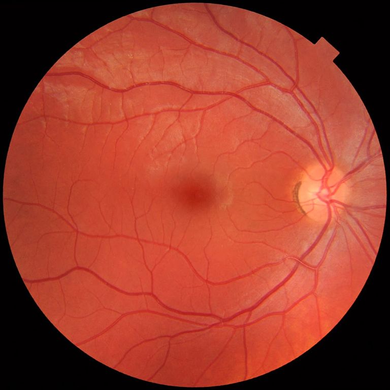
Used with permission., CC0, via Wikimedia Commons.
Many remember rods and cones from middle school class. Each type of photoreceptor cell has a specific function. Rods are more numerous (more than 120 million) and are more sensitive than cones. Cones, number 6 million, and see the colors red, blue, and green.
Vitreous
The vitreous fluid or vitreous humor is a clear, gel-like fluid that fills the back of the eye from the lens to the retina. The vitreous humor is composed of mostly water with a small percentage of collagen, glycosaminoglycans (sugars), electrolytes (salts), and proteins. The function of the vitreous humor is to hold the structure of the eye together while allowing light to pass through to the retina.
With aging, or sometimes earlier in patients with high glasses prescription, the vitreous humor may shrink or degenerate. The degeneration may cause vitreous floaters that appear as spots, lines, or rings. Floaters are usually not harmful unless the vitreous pulls the retina resulting in a tear called a retinal detachment. All new floaters, flashes of light, or a new curtain-like effect appearing in your vision should be checked immediately by an eye doctor. They will dilate your eye and check the retina a full 360°.
Optic nerve
The optic nerve is in the very back of the eye and is an extension of the brain into the eye. The optic nerve is comprised of 1 million nerve fibers that carry signals from the eye to the brain. The optic nerve is the reason for the “blind spot” when driving.
The optic nerve can be affected by many eye diseases and pathologies, both genetic and inherited. Most common conditions include glaucoma, optic neuritis, and optic nerve atrophy.
Glaucoma is a painless, vascular condition of the optic nerve that affect peripheral vision in 2.7 million Americans over 40. Glaucoma cannot be diagnosed without an eye exam and is the primary reason you should have your eyes checked annually even if you have 20/20 vision.
Choroid
The choroid is a layer of blood vessels that sits underneath and supplies the outer retina with nutrients and maintains the temperature and volume of the eye. The choroidal blood circulation is 85% of the total blood flow in the eye.
The blood vessels in the choroid are affected by wet age-related macular degeneration (ARMD). Wet ARMD is less common than the dry form, but results in much more vision loss. In ARMD, due to a combination of genetic and environmental factors, new blood vessels form from the choroid and break into the error resulting in bleeding in the eye (neovascularization).
The eye is a complicated organ, and each tiny part is necessary for clear vision.
- Your eyes are capable of processing 36,000 pieces of information per hour.
- Your iris has 256 unique characteristics, far more than your fingerprints (40).
- Your eyes are some of the fastest and strongest muscles in your body.
More interesting facts about your eyes can be found here!